
NCERT Solutions for Class 11th BIOLOGY
Chapter 18 Body Fluids And Ciculation
1. Name the components of the formed elements in the blood and mention one major function of each of them.
Solutlion: Blood corpuscles are the formed ele-ments in the blood, they constitute 45% of the blood. Formed elements are – (erythrocytes, RBCs or red blood corpuscles), (leucocytes, WBCs or white blood corpuscles) and throm¬bocytes or blood platelets. The major function of RBCs is to transport oxygen from lungs to body tissues and COz from body tissues to the lungs. White blood cells provide immunity to the body. Blood platelets play important role in blood clotting.
Solutlion: Blood corpuscles are the formed ele-ments in the blood, they constitute 45% of the blood. Formed elements are – (erythrocytes, RBCs or red blood corpuscles), (leucocytes, WBCs or white blood corpuscles) and throm¬bocytes or blood platelets. The major function of RBCs is to transport oxygen from lungs to body tissues and COz from body tissues to the lungs. White blood cells provide immunity to the body. Blood platelets play important role in blood clotting.
2. What is the importance of plasma proteins?
Solutlion: Plasma proteins constitute about 7 to 8% of plasma. These mainly include albumin, globulin, prothrombin and fibrinogen. Prothrombin and fibrinogen are needed for blood clotting. Albumins and globulins retain water in blood plasma and helps in maintainingosmoticbalance. Certain globulins
Solutlion: Plasma proteins constitute about 7 to 8% of plasma. These mainly include albumin, globulin, prothrombin and fibrinogen. Prothrombin and fibrinogen are needed for blood clotting. Albumins and globulins retain water in blood plasma and helps in maintainingosmoticbalance. Certain globulins
3. Match Column I with Column II.
Column I Column II
(a) Eosinophils (i) Coagulation
(b) RBC (ii) Universal recipient
(c) AB Group (iii) Resist infections
(d) Platelets (iv) Contraction of heart
(e) Systol (v) Gas transport
Solutlion.(a) – (iii); (b) – (v); (c) – (ii); (d) – (i); (e) – (iv).
Column I Column II
(a) Eosinophils (i) Coagulation
(b) RBC (ii) Universal recipient
(c) AB Group (iii) Resist infections
(d) Platelets (iv) Contraction of heart
(e) Systol (v) Gas transport
Solutlion.(a) – (iii); (b) – (v); (c) – (ii); (d) – (i); (e) – (iv).
4. Why do we consider blood as a connective tissue?
Solutlion: A connective tissue connects different tissues or organs of the body. It consists of living cells and extracellular matrix. Blood is vascular connective tissue, it is a mobile tissue consisting of fluid matrix and free cells. Blood transports materials from one place to the other and thereby establishes connectivity between different body parts.
Solutlion: A connective tissue connects different tissues or organs of the body. It consists of living cells and extracellular matrix. Blood is vascular connective tissue, it is a mobile tissue consisting of fluid matrix and free cells. Blood transports materials from one place to the other and thereby establishes connectivity between different body parts.
5. What is the difference between lymph and blood?
Solutlion: The differences between blood and lymph are given below:
 Class 11 BIOLOGY SOLUTION
Solutlion: The differences between blood and lymph are given below:
Class 11 BIOLOGY SOLUTION
6. What is meant by double circulation? What is its significance?
Solutlion: The type of blood circulation in which oxygenated blood and deoxygenated blood do not get mixed is termed double circulation. It includes systemic circulation and pulmonary circulation. The circulatory pathway of double circulation is given in the following flow chart.
 Class 11 BIOLOGY SOLUTION
Flow chart: Double blood circulation Double circulation or separation of systemic and pulmonary circulations provides a higher metabolic rate to the body and also allows the two circulations to have different blood pressures according to the need of the organs they supply.
Solutlion: The type of blood circulation in which oxygenated blood and deoxygenated blood do not get mixed is termed double circulation. It includes systemic circulation and pulmonary circulation. The circulatory pathway of double circulation is given in the following flow chart.
Class 11 BIOLOGY SOLUTIONFlow chart: Double blood circulation Double circulation or separation of systemic and pulmonary circulations provides a higher metabolic rate to the body and also allows the two circulations to have different blood pressures according to the need of the organs they supply.
Question 7: Write the differences between:
(a) Blood and Lymph
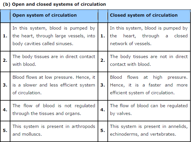
(b) Open and Closed system of circulation
(c) Systole and Diastole
(d) P-wave and T-wave
Answer


(a) Blood and Lymph
(b) Open and Closed system of circulation
(c) Systole and Diastole
(d) P-wave and T-wave
Answer
Question 8: Describe the evolutionary change in the pattern of heart among the vertebrates.
Answer All vertebrates possess a heart – a hollow muscular organ composed of cardiac muscle fibres. The function of the heart is to pump oxygen to all parts of the body. The evolution of the heart is based on the separation of oxygenated blood from deoxygenated blood for efficient oxygen transport.
In fishes, the heart was like a hollow tube. This evolved into the four-chambered heart in mammals. Piscean heart
Fish has only two chambers in its heart – one auricle and one ventricle. Since both the auricle and the ventricle remain undivided, only deoxygenated blood passes through it. The deoxygenated blood enters the gills for oxygenation from the ventricle. It has additional chambers such as sinus venosus and conus arteriosus.

Amphibian heart
Amphibians, such as frogs, have three-chambered hearts, with two auricles and one ventricle. The auricle is divided into a right and a left chamber by an inter-auricular septum, while the ventricle remains undivided. Additional chambers such as sinus venosus and conus arteriosus are also present. The oxygenated blood from the lungs enters the left auricle and simultaneously, the deoxygenated blood from the body enters the right auricle. Both these auricles empty into the ventricle, wherein the oxygenated and deoxygenated blood get mixed to some extent.

Reptilian heart
Reptiles have incomplete four-chambered hearts, except for crocodiles, alligators, and gharials. They have only one accessory chamber called sinus venosus. The reptilian heart also shows mixed blood circulation.

Avian and mammalian hearts
They have two pairs of chambers for separating oxygenated and deoxygenated bloods. The heart is divided into four chambers. The upper two chambers are called atria and the lower two chambers are called ventricles. The chambers are separated by a muscular wall that prevents the mixing of the blood rich in oxygen with the blood rich in carbon dioxide.
 Class 11 BIOLOGY SOLUTION
Answer All vertebrates possess a heart – a hollow muscular organ composed of cardiac muscle fibres. The function of the heart is to pump oxygen to all parts of the body. The evolution of the heart is based on the separation of oxygenated blood from deoxygenated blood for efficient oxygen transport.
In fishes, the heart was like a hollow tube. This evolved into the four-chambered heart in mammals. Piscean heart
Fish has only two chambers in its heart – one auricle and one ventricle. Since both the auricle and the ventricle remain undivided, only deoxygenated blood passes through it. The deoxygenated blood enters the gills for oxygenation from the ventricle. It has additional chambers such as sinus venosus and conus arteriosus.
Amphibian heart
Amphibians, such as frogs, have three-chambered hearts, with two auricles and one ventricle. The auricle is divided into a right and a left chamber by an inter-auricular septum, while the ventricle remains undivided. Additional chambers such as sinus venosus and conus arteriosus are also present. The oxygenated blood from the lungs enters the left auricle and simultaneously, the deoxygenated blood from the body enters the right auricle. Both these auricles empty into the ventricle, wherein the oxygenated and deoxygenated blood get mixed to some extent.
Reptilian heart
Reptiles have incomplete four-chambered hearts, except for crocodiles, alligators, and gharials. They have only one accessory chamber called sinus venosus. The reptilian heart also shows mixed blood circulation.
Avian and mammalian hearts
They have two pairs of chambers for separating oxygenated and deoxygenated bloods. The heart is divided into four chambers. The upper two chambers are called atria and the lower two chambers are called ventricles. The chambers are separated by a muscular wall that prevents the mixing of the blood rich in oxygen with the blood rich in carbon dioxide.
Class 11 BIOLOGY SOLUTION
Question 9: Why do we call our heart myogenic?
Answer In the human heart, contraction is initiated by a special modified heart muscle known as sinoatrial node. It is located in the right atrium. The SA node has the inherentpower of generating a wave of contraction and controlling the heart beat. Hence, it is known as the pacemaker. Since the heart beat is initiated by the SA node and the impulse of contraction originates in the heart itself, the human heart is termed myogenic. The hearts of vertebrates and molluscs are also myogenic.
Answer In the human heart, contraction is initiated by a special modified heart muscle known as sinoatrial node. It is located in the right atrium. The SA node has the inherentpower of generating a wave of contraction and controlling the heart beat. Hence, it is known as the pacemaker. Since the heart beat is initiated by the SA node and the impulse of contraction originates in the heart itself, the human heart is termed myogenic. The hearts of vertebrates and molluscs are also myogenic.
10. Sino-atrial node is called the pacemaker of our heart. Why?
Solutlion: Sino-atrial node (SAN) is a mass of neuromuscular tissue which lies in the wall of right atrium. It is responsible for initiating and maintaining the rhythmic contractile activity of the heart. Therefore, it is called the pacemaker.
Solutlion: Sino-atrial node (SAN) is a mass of neuromuscular tissue which lies in the wall of right atrium. It is responsible for initiating and maintaining the rhythmic contractile activity of the heart. Therefore, it is called the pacemaker.
11. What is the significance of atrio-ventricular node and atrio-ventricular bundle in the functioning of heart?
Solutlion: Atrio-ventricular node (AVN) is a mass of neuromuscular tissue, which is situated in wall of. right atrium, near the base of inter-atrial septum. AV node is the pacesetter of the heart,- as it transmits the impulses initiated by SA node to all parts of ventricles. Atrio-ventricular bundle (A-V bundle) or bundle of His is a mass of specialised fibres which originates from the AVN. Within the myocardium of the ventricles the branches of bundle of His divide into a network of fine fibres called Purkinje fibres. The bundle of His and the Purkinje fibres convey impulse of contraction from the AVN to the myocardium of the ventricles.
Solutlion: Atrio-ventricular node (AVN) is a mass of neuromuscular tissue, which is situated in wall of. right atrium, near the base of inter-atrial septum. AV node is the pacesetter of the heart,- as it transmits the impulses initiated by SA node to all parts of ventricles. Atrio-ventricular bundle (A-V bundle) or bundle of His is a mass of specialised fibres which originates from the AVN. Within the myocardium of the ventricles the branches of bundle of His divide into a network of fine fibres called Purkinje fibres. The bundle of His and the Purkinje fibres convey impulse of contraction from the AVN to the myocardium of the ventricles.
12. Define a cardiac cycle and the cardiac output.
Solutlion: The sequential events in the heart which are repeated cyclically is called cardiac cycle and it consists of systole (contraction) and diastole (relaxation) of both the atria and ventricles. The duration of a cardiac cycle is 0.8 seconds. Periods of cardiac cycle are atrial systole (0.1 second), ventricular systole (0.3 second) and complete cardiac diastole (0.4 second).
The amount of blood pumped by heart per minute is called cardiac output. It is calculated by multiplying stroke volume (volume of blood pumped by each ventricle per minute) with heart rate (number of beats per minute). The heart of normal person beats 72 times per minute and pumps out about 70 mL of blood per beat. Therefore, cardiac output averages 5000 mL or 5 litres.
13. Explain heart sounds.
Solutlion: The beating of heart produces characteristic sounds which can be heard by using stethoscope. In a normal person, two sounds are produced per heart beat. The first heart sound Tubb’ is low pitched, not very loud and of long duration. It is caused partly by the closure of the bicuspid and tricuspid valves and partly by the contraction of muscles in the ventricles.
The second heart sound ‘dubb’ is high pitched, louder, sharper and shorter in duration. It is caused by the closure of the semilunar valves and marks the end of ventricular systole.
Question 14: Draw a standard ECG and explain the different segments in it.
Answer Electrocardiogram is a graphical representation of the cardiac cycle produced by an electrograph. The diagrammatic representation of a standard ECG is shown below.

A typical human electrocardiogram has five waves – P, Q, R, S, and T. The P, R, and T-waves are above the base line and are known as positive waves. The Q and S-waves are below the base line and are known as negative waves. The P-wave is of atrial origin, while the Q, R, S, and T-waves are of ventricular origin.
(a) The P-wave indicates atrial depolarisation. During this wave, the impulse of contraction is generated by the SA node. The PQ-wave represents atrial contraction.
(b) The QR-wave is preceded by ventricular contraction. It represents the spread of the impulse of contraction from the AV node to the wall of the ventricle. It leads to ventricular depolarisation.
(c) The RS-wave represents ventricular contraction of about 0.3 sec.
(d) The ST-wave represents ventricular relaxation of about 0.4 sec. During this phase, the ventricles relax and return to their normal state.
(e) The T-wave represents ventricular relaxation.
Answer Electrocardiogram is a graphical representation of the cardiac cycle produced by an electrograph. The diagrammatic representation of a standard ECG is shown below.
A typical human electrocardiogram has five waves – P, Q, R, S, and T. The P, R, and T-waves are above the base line and are known as positive waves. The Q and S-waves are below the base line and are known as negative waves. The P-wave is of atrial origin, while the Q, R, S, and T-waves are of ventricular origin.
(a) The P-wave indicates atrial depolarisation. During this wave, the impulse of contraction is generated by the SA node. The PQ-wave represents atrial contraction.
(b) The QR-wave is preceded by ventricular contraction. It represents the spread of the impulse of contraction from the AV node to the wall of the ventricle. It leads to ventricular depolarisation.
(c) The RS-wave represents ventricular contraction of about 0.3 sec.
(d) The ST-wave represents ventricular relaxation of about 0.4 sec. During this phase, the ventricles relax and return to their normal state.
(e) The T-wave represents ventricular relaxation.
Comments
Post a Comment