
NCERT Solutions for Class 11th Biology
Chapter 7 Structural Organisation in Animals
Question 1: Answer in one word or one line.
(i) Give the common name of Periplaneta americana.
(ii) How many spermathecae are found in earthworm?
(iii) What is the position of ovaries in the cockroach?
(iv) How many segments are present in the abdomen of cockroach?
(v) Where do you find malphigian tubules?
Answer (i) The common name of Periplaneta americana is the American cockroach.
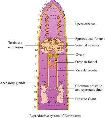
(ii) Four pairs of spermathecae are present in earthworms. They are located between sixth and the ninth segments. They help in receiving and storing the spermatozoa during copulation.
(iii) In a cockroach, the pair of ovaries is located between 12th and 13th abdominal segments.
(iv) In both sexes, the abdomen of a cockroach consists of ten segments.
(v) Malphigian tubules are main excretory organs of cockroaches. They form a part of the alimentary canal.
(i) Give the common name of Periplaneta americana.
(ii) How many spermathecae are found in earthworm?
(iii) What is the position of ovaries in the cockroach?
(iv) How many segments are present in the abdomen of cockroach?
(v) Where do you find malphigian tubules?
Answer (i) The common name of Periplaneta americana is the American cockroach.
(ii) Four pairs of spermathecae are present in earthworms. They are located between sixth and the ninth segments. They help in receiving and storing the spermatozoa during copulation.
(iii) In a cockroach, the pair of ovaries is located between 12th and 13th abdominal segments.
(iv) In both sexes, the abdomen of a cockroach consists of ten segments.
(v) Malphigian tubules are main excretory organs of cockroaches. They form a part of the alimentary canal.
Question 2: Answer the following:
(i) What is the function of nephridia?
(ii) How many types of nephridia are found in earthworm based on their location?
Answer (i) Nephridia are segmentally arranged excretory organs present in earthworms.
(ii) On the basis of their location, three types of nephridia are found in earthworms. They are:
(a) Septal nephridia: These are present on both sides of the inter-segmental septa behind the 15th segment. They open into the intestines.
(b) Integumentary nephridia: These lie attached to the body wall from the third segment to the last segment, which opens on the body surface.
(c) Pharyngeal nephridia: These are present as three paired tufts in fourth, fifth, and sixth segments.
Question 3: Draw a labelled diagram of the reproductive organs of an earthworm.
Question 4: Draw a labelled diagram of alimentary canal of a cockroach.
Answer

Answer
5. Distinguish between the following:
(a) Prostomium and peristomium
(b) Septal nephridium and pharyngeal
Solution: (a) Differences between prostomium and peristomium are


(b) Differences between septal and pharyngeal nephridia are:
 Download our app
(a) Prostomium and peristomium
(b) Septal nephridium and pharyngeal
Solution: (a) Differences between prostomium and peristomium are
(b) Differences between septal and pharyngeal nephridia are:
Download our app
6. What are the cellular components of blood?
Solution: Blood is a fluid connective tissue. It is composed of plasma (fluid) and blood cells (corpuscles). Cellular components of blood (blood corpuscles) constitute about 45% of blood volume.
Three types of blood cells are:
(i) Erythrocytes or red blood cells: They are most abundant blood cells. Normal RBC count is 5-5.5 million/mm3 in males and 4.5-5 million/mm3 in females) RBCs help in transport of gases and maintain blood pH.
(ii) Leucocytes or white blood cells: The normal WBC count is 5000-6000/mm3 of blood. They are involved in immune response of body and act as soldiers and scavangers.
(iii) Thrombocytes or blood platelets: There are about 2,50,000 platelets/mm3 of blood. They are involved in blood clotting.
8. Mark the odd one in each series.
(a) Areolar tissue; blood; neuron; tendon
(b) RBC; WBC; platelets; cartilage
(c) Exocrine; endocrine; salivary gland; ligament
(d) Maxilla; mandible; labrum; antennae
(e) Protonema; mesothorax; metathorax; coxa.
Solution:
(a) Neuron: Areolar tissue, blood and tendon are connective tissues while neuron is a part a nervous tissue.
(b) Cartilage: RBC, WBC and platelets are parts of vascular connective tissue while cartilage is skeletal connective tissue.
(c) Ligament: Ligament is a connective tissue.
(d) Antennae: Maxilla, mandible and labrum are mouth parts of cockroach while antennae are sense organs.
(e) Protonema: Protonema is a filamentous juvenile stage in life cycle of Bryophytes, while mesothorax, metathorax and coxa are appendages of cockroach.
9. Match the terms in column I with those in column II.
Column I
|
Column II
|
| (a) Compound epithelium (b) Compound eye (c) Septal nephridia (d) Open circulatory system (e) Typhlosole (f) Osteocytes (g) Genitalia | (i) Alimentry canal (ii) Cockroach (iii) Skin (iv) Mosaic vision (v) Earthworm (vi) Phallomere (vii) Bone |
Solution: (a) – (iii), (b) – (iv), (c) – (v), (d) – (ii), (e) – (i), (f) – (vii), (g) – (vi)
11. Describe various types of epithelial tissues with the help of labelled diagrams.
Solution: Epithelial tissue is a tissue made of one or more layers of compactly arranged cells that covers external surface and internal free surface of body organs and which is underlined by a basement membrane. The various types of epithelial tissue along with the diagram are given below:
(i) Simple epithelium : It is composed of single layer of cells which rest on basement membrane. Simple epithelium generally occurs over secretory and absorptive surfaces and forms lining of body cavities, ducts and tubes. Simple epithelium is of several types.
(a) Squamous epithelium: It consists of single layer of flat cells, tightly linked together and have centrally located oval or spherical nucleus. It is also called pavement epithelium. It is found in walls of blood vessels, air sacs of lungs, and lining of eye lens.
(b) Cuboidal epithelium: Cells of cuboidal epithelium are as tall as wide, with centrally placed nucleus. Its main functions are secretion and absorption. It lines sweat gland, thyroid follicles, salivary glands. Brush bordered cuboidal epithelium, i.e., cells having microvilli on their free surface lines proximal part of uriniferous tubule, pancreatic duct, testis and ovary.
(c) Columnar epithelium: Cells are with basally located nucleus. It helps in secretion and absorption. It occurs in lining of intestine, stomach, gall bladder.
(d) Ciliated epithelium: Free surface of columnar and cuboidal cells are covered with cilia. Cilia help in moving fluids, particles, mucus, etc. in a specific direction. It occurs in the inner surface of Fallopian tubules, nasal passage, bronchioles.

(e) Pseudostratified epithelium: It consists of single layer of cells but some cells are shorter than others. Due to difference in size of cells, the epithelium appears 2-3 layered. Pseudostratified columnar epithelium occurs in urethra and parotid salivary gland. Pseudostratified columnar ciliated epithelium (only larger cells ciliated) occurs in lining layer of nasal’ chambers, trachea and large bronchi. It helps in moving mucus and foreign particles.

(ii) Compoundepithelium/stratifiedepithelium: It is multilayered epithelium where cells of only the lowermost or basal layer are in contact with basement membrane. It provides protection against mechanical and chemical stresses and has limited role in secretion and absorption. It covers dry surface of skin, moist surface of buccal cavity, pharynx, etc. Different types of compound epithelium are:
(a) Stratified squamous epithelium: The cells of outer layer are flattened and squamous while the inner layers are cuboidal cells. It is of two types: Non- keratinised lining oesophagus, pharynx, buccal cavity, cornea, vagina and anal canal and keratinised (comified): forming epidermis of skin, hair, horn and nail.
 Download our app
(b) Stratified cuboidal epithelium: The outer layer of cuboidal cells and basal layer of columnar cells. It lines ducts of sweat glands, large salivary and pancreatic ducts.
(c) Stratified columnar epithelium: Both upper and basal layers are made of columnar cells, e.g., epiglottis covering, part of urethra.
(d) Stratified ciliated columnar epithelium: Outer layer consists of ciliated columnar cells and basal layer of columnar cells, e.g., larynx.
(iii) Transitional Epithelium: This is stratified epithelium which contains cuboidal or columnar shaped cells, which are thin and stretchable. No basement membrane is present as it would impede stretchability. It lines the inner surface of renal calyces, urinary bladder, ureter. Because of its t distribution, it is also called urothelium.

(iv) Glandular epithelium: It consists of specialised epithelial cells which synthesise intracellular macromolecules (protein in pancreas, lipids in adrenal glands, glycoprotein in salivary glands and all the three in mammary glands) and pour out the same in the form of a useful fluid secretion which is different from blood or any other extracellular fluid. Glands can be unicellular or multicellular on the basis of number of cells.
(a) Unicellular glands: Single-celled, e.g., goblet (mucous) cells of respiratory tract and alimentary canal.
(b) Multicellular glands: Consist of cluster of cells, e.g., Salivary glands.

On the basis of presence or absence of duct glands can be:
(a) Exocrine glands : These glands pour their secretion through a duct. They secrete milk, saliva, mucus, earwax. e.g., goblet cells, salivary glands, tear glands, gastric glands, intestinal glands.
(b) Endocrine glands: They are ductless glands, which pour their secretions into blood or lymph for reaching the target region. Their secretion is called hormone e.g., pituitary gland, thyroid gland, parathyroid glands, adrenal glands.
(c) Heterocrine glands: Both exocrine and endocrine, e.g., pancreas.
On basis of mode of secretion glands can be:
(a) Merocrine: Secretion is discharged
through diffusion, e g., goblet cells, sweat glands.
(b) Apocrine glands: Glandular secretion accumulates in the terminal part of the cell which is pinched off, e.g., mammary glands.
(c) Holocrine glands : The cell filled with secretory product disintegrates during discharge of the product, e.g., sebaceous gland.
(v) Modified epithelium : It is of following types:
(a) Germinal epithelium (generally cuboidal, produces gametes), (b) Glandular epithelium (columnar or cuboidal secretes chemicals and mucus), (c) Sensory epithelium or neuroepithelium. Epithelial cells having sensory hair on free surface and connected with nerve fibres on the other surface (generally columnar, receives and conveys stimuli), e.g, nasal epithelium, taste buds, retina, sensory spots of internal ear. (d) Pigmented epithelium – The cells possess melanin granules, e.g, retinal layer in contact with choroid of eye.
Solution: Epithelial tissue is a tissue made of one or more layers of compactly arranged cells that covers external surface and internal free surface of body organs and which is underlined by a basement membrane. The various types of epithelial tissue along with the diagram are given below:
(i) Simple epithelium : It is composed of single layer of cells which rest on basement membrane. Simple epithelium generally occurs over secretory and absorptive surfaces and forms lining of body cavities, ducts and tubes. Simple epithelium is of several types.
(a) Squamous epithelium: It consists of single layer of flat cells, tightly linked together and have centrally located oval or spherical nucleus. It is also called pavement epithelium. It is found in walls of blood vessels, air sacs of lungs, and lining of eye lens.
(b) Cuboidal epithelium: Cells of cuboidal epithelium are as tall as wide, with centrally placed nucleus. Its main functions are secretion and absorption. It lines sweat gland, thyroid follicles, salivary glands. Brush bordered cuboidal epithelium, i.e., cells having microvilli on their free surface lines proximal part of uriniferous tubule, pancreatic duct, testis and ovary.
(c) Columnar epithelium: Cells are with basally located nucleus. It helps in secretion and absorption. It occurs in lining of intestine, stomach, gall bladder.
(d) Ciliated epithelium: Free surface of columnar and cuboidal cells are covered with cilia. Cilia help in moving fluids, particles, mucus, etc. in a specific direction. It occurs in the inner surface of Fallopian tubules, nasal passage, bronchioles.
(e) Pseudostratified epithelium: It consists of single layer of cells but some cells are shorter than others. Due to difference in size of cells, the epithelium appears 2-3 layered. Pseudostratified columnar epithelium occurs in urethra and parotid salivary gland. Pseudostratified columnar ciliated epithelium (only larger cells ciliated) occurs in lining layer of nasal’ chambers, trachea and large bronchi. It helps in moving mucus and foreign particles.
(ii) Compoundepithelium/stratifiedepithelium: It is multilayered epithelium where cells of only the lowermost or basal layer are in contact with basement membrane. It provides protection against mechanical and chemical stresses and has limited role in secretion and absorption. It covers dry surface of skin, moist surface of buccal cavity, pharynx, etc. Different types of compound epithelium are:
(a) Stratified squamous epithelium: The cells of outer layer are flattened and squamous while the inner layers are cuboidal cells. It is of two types: Non- keratinised lining oesophagus, pharynx, buccal cavity, cornea, vagina and anal canal and keratinised (comified): forming epidermis of skin, hair, horn and nail.
Download our app(b) Stratified cuboidal epithelium: The outer layer of cuboidal cells and basal layer of columnar cells. It lines ducts of sweat glands, large salivary and pancreatic ducts.
(c) Stratified columnar epithelium: Both upper and basal layers are made of columnar cells, e.g., epiglottis covering, part of urethra.
(d) Stratified ciliated columnar epithelium: Outer layer consists of ciliated columnar cells and basal layer of columnar cells, e.g., larynx.
(iii) Transitional Epithelium: This is stratified epithelium which contains cuboidal or columnar shaped cells, which are thin and stretchable. No basement membrane is present as it would impede stretchability. It lines the inner surface of renal calyces, urinary bladder, ureter. Because of its t distribution, it is also called urothelium.
(iv) Glandular epithelium: It consists of specialised epithelial cells which synthesise intracellular macromolecules (protein in pancreas, lipids in adrenal glands, glycoprotein in salivary glands and all the three in mammary glands) and pour out the same in the form of a useful fluid secretion which is different from blood or any other extracellular fluid. Glands can be unicellular or multicellular on the basis of number of cells.
(a) Unicellular glands: Single-celled, e.g., goblet (mucous) cells of respiratory tract and alimentary canal.
(b) Multicellular glands: Consist of cluster of cells, e.g., Salivary glands.
On the basis of presence or absence of duct glands can be:
(a) Exocrine glands : These glands pour their secretion through a duct. They secrete milk, saliva, mucus, earwax. e.g., goblet cells, salivary glands, tear glands, gastric glands, intestinal glands.
(b) Endocrine glands: They are ductless glands, which pour their secretions into blood or lymph for reaching the target region. Their secretion is called hormone e.g., pituitary gland, thyroid gland, parathyroid glands, adrenal glands.
(c) Heterocrine glands: Both exocrine and endocrine, e.g., pancreas.
On basis of mode of secretion glands can be:
(a) Merocrine: Secretion is discharged
through diffusion, e g., goblet cells, sweat glands.
(b) Apocrine glands: Glandular secretion accumulates in the terminal part of the cell which is pinched off, e.g., mammary glands.
(c) Holocrine glands : The cell filled with secretory product disintegrates during discharge of the product, e.g., sebaceous gland.
(v) Modified epithelium : It is of following types:
(a) Germinal epithelium (generally cuboidal, produces gametes), (b) Glandular epithelium (columnar or cuboidal secretes chemicals and mucus), (c) Sensory epithelium or neuroepithelium. Epithelial cells having sensory hair on free surface and connected with nerve fibres on the other surface (generally columnar, receives and conveys stimuli), e.g, nasal epithelium, taste buds, retina, sensory spots of internal ear. (d) Pigmented epithelium – The cells possess melanin granules, e.g, retinal layer in contact with choroid of eye.
12. Distinguish between
(a) Simple epithelium and compound epithelium.
(b) Cardiac muscle and striated muscle.
(c) Dense regular and dense irregular connective tissues.
(d) Adipose and blood tissue.
(e) Simple gland and compound gland.
Solution:
(a) Differences between simple and compound epithelium are as follows:
 Download our app
(b) Differences between cardiac and striated muscles are as follows:


(c) Differences between dense regular and dense irregular connective tissues are as follows:

(d) Differences between adipose tissue and blood tissue are as follows:

(e) Differences between simple gland and compound gland are as follows:

(a) Simple epithelium and compound epithelium.
(b) Cardiac muscle and striated muscle.
(c) Dense regular and dense irregular connective tissues.
(d) Adipose and blood tissue.
(e) Simple gland and compound gland.
Solution:
(a) Differences between simple and compound epithelium are as follows:
Download our app(b) Differences between cardiac and striated muscles are as follows:
(c) Differences between dense regular and dense irregular connective tissues are as follows:
(d) Differences between adipose tissue and blood tissue are as follows:
(e) Differences between simple gland and compound gland are as follows:
13. Draw a neat diagram of digestive system of frog.
Solution:

Solution:
Question 14: Mention the function of the following
(a) Ureters in frog
(b) Malpighian tubules
(c) Body wall in earthworm
Answer (a) Ureters in frogs: A ureter acts as a urinogenital duct, which carries sperms along with urine in male frogs.
(b) Malphigian tubules: Malphigian tubules are excretory organs in cockroaches.
(c) Body wall in earthworms: In earthworms, the body wall consists of muscle layers. It helps in movement and burrowing.
Comments
Post a Comment